Artsy Lumbar Vertebrae Model with Sacrum, Coccyx & Herniated Disc – Life-Size, High-Precision Anatomy Tool
Where Anatomy Meets Clarity – Teach, Demonstrate, and Empower with Confidence
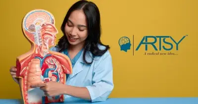
The Artsy Lumbar Vertebrae with Sacrum, Coccyx & Herniated Disc Model brings the intricacies of the human lower spine to life. Designed for orthopedic professionals, physiotherapists, chiropractors, anatomy educators, and students, this life-size model offers a remarkably detailed and tactile view of the lumbosacral region.
Featuring five lumbar vertebrae (L1–L5), intervertebral discs, sacrum, coccyx, and realistic yellow spinal nerves, it provides a comprehensive anatomical overview. A standout feature is the raised, vividly colored herniated disc at L4-L5, giving users a powerful tool to explain disc pathology, nerve impingement, and sciatic pain with unmatched visual impact.
Whether used in anatomy classrooms, rehab centers, consultation rooms, or mobile clinics, this model is compact yet rich in detail—transforming spinal education from theory into tangible understanding.
🔍 Key Features:
🦴 Complete Lumbosacral Segment
Includes L1–L5 vertebrae, intervertebral discs, sacrum, and coccyx
Ideal for showing spinal curvature, nerve pathways, and pelvic transition
Supported by a stable steel-mounted base for 360° demonstration
📛 Realistic Herniated Disc at L4–L5
Textured, red-highlighted protrusion simulates disc bulge with accuracy
Clearly shows how herniation affects adjacent spinal nerve roots
Perfect for explaining common issues like sciatica and lower back pain
🧠 Educational Utility Across Disciplines
For orthopedic consultations, chiropractic sessions, and physiotherapy evaluations
Excellent teaching tool for BPT, MBBS, and allied health anatomy curricula
Students can palpate vertebral structures, understand spinal mechanics, and memorize landmarks efficiently
🧳 Compact Yet Clinically Insightful
Lightweight and easy to carry for workshops, mobile demos, or campus rounds
Designed for daily clinical or academic use—resistant to stains, chips, and deformation
PVC-based synthetic resin ensures lasting quality and safety
💡 Ideal For:
Orthopedic and neuro consultations – visually explain lumbar disc issues
Rehabilitation and pain clinics – support patient compliance through clarity
Medical and nursing education – offer students a 3D tactile learning experience
Postural education sessions – reinforce understanding of spinal support and flexibility
“It’s helped me explain disc issues to patients far more effectively. They understand it the moment they see it.”
— Dr. Kamal V., Orthopedic Consultant, Delhi
🎯 Why Choose the Artsy Herniated Disc Lumbar Spine Model?
✔ Life-size and anatomically precise
✔ Clearly marked spinal nerves and foramina
✔ Visualizes the transition from spine to pelvis
✔ Easy to transport, easy to understand, hard to forget
From Classroom to Clinic – A Tool That Connects
The Artsy Lumbar Vertebrae with Herniated Disc Model isn’t just an anatomical prop—it’s a daily-use asset that improves communication, learning, and decision-making. Its compact size fits any space, but its educational power is immense. When explaining spinal compression, disc degeneration, or nerve root impingement, no diagram or scan compares to the hands-on clarity this model offers.
From orthopedic surgeons and physiotherapists to anatomy professors and patient educators, this tool is trusted by professionals who value simplicity, accuracy, and impact.
“It’s one of the most used models in our training center. Explaining back pain, posture, or disc injuries becomes far easier with this on hand.”
— Dr. Lee M., Clinical Educator, Singapore
Specifications at a Glance:
Model Includes: L1–L5 vertebrae, herniated disc, sacrum, coccyx, spinal nerves
Material: Medical-grade, non-toxic PVC
Mounting: Fixed on a stable steel rod and base
Size: Life-size dimensions for anatomical accuracy
Weight: Lightweight and portable
A Compact Tool With Maximum Educational Impact
Artsy's life-size lumbar spine model demonstrates more than just anatomy—it builds trust, clarifies complexity, and improves outcomes. Whether you're guiding a patient through their condition or helping a student prepare for exams, this model delivers learning that lasts.