Description
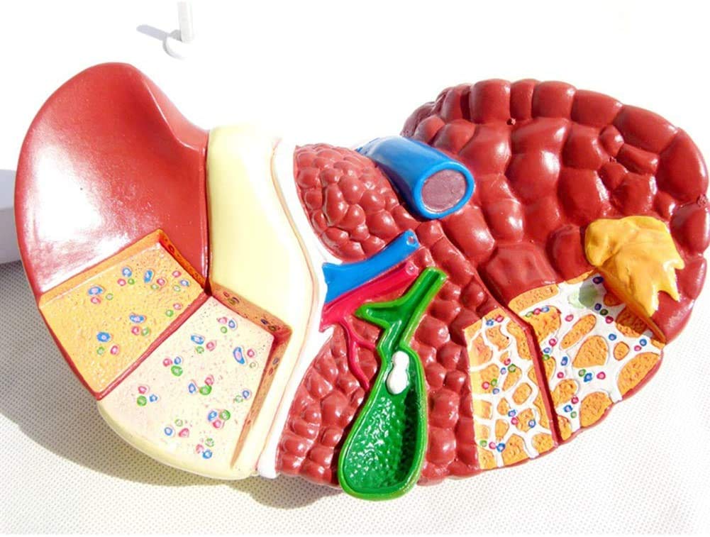
Introducing the Diseased Liver (Cancer) Anatomy Model, a highly detailed anatomical model designed to provide an in-depth understanding of liver anatomy and the effects of liver cancer. Ideal for students, educators, hepatologists, oncologists, and medical professionals, this model is an essential educational tool for studying and teaching the complex pathology of liver cancer.
Key Features:
-
Detailed Anatomy: The model accurately represents the human liver, showcasing its lobes, vessels, and internal structures. It provides a clear view of the liver’s anatomical features, including the hepatic artery, portal vein, and bile ducts.
-
Pathological Conditions: The model includes detailed representations of liver cancer and related pathologies, such as:
- Hepatocellular carcinoma (HCC)
- Metastatic liver cancer
- Cirrhosis-related changes
- Liver nodules and lesions
-
Realistic Representation: Crafted to mimic the appearance and texture of real liver tissue, the model features lifelike details that enhance the learning experience. The realistic design helps users visualize and understand the anatomical and pathological changes associated with liver cancer.
-
Cross-Sectional Views: The model includes cross-sectional views that provide an in-depth look at the internal structure of the liver and the progression of cancerous growths. This design allows for a deeper understanding of how liver cancer affects the organ’s structure and function.
-
High-Quality Materials: Made from durable, high-quality materials, this model is built to withstand frequent handling. The robust construction ensures longevity and provides reliable educational value over time.
-
Educational Labels: The model includes clearly marked labels on key anatomical features and pathological conditions, making it easy to identify and learn about various parts of the liver and the impact of cancer. An accompanying guide provides detailed descriptions and explanations of each labeled structure and pathology.
-
Versatile Use: Suitable for a wide range of applications, including medical training, biology and anatomy classes, oncology education, and as a display piece in clinics and healthcare facilities.
Benefits:
- Enhanced Learning: Provides a hands-on, interactive way to study and understand liver anatomy and the effects of liver cancer, aiding in the retention of complex information.
- Improved Teaching: Serves as a valuable visual aid for educators, helping to make lessons more engaging and informative.
- Professional Use: Assists healthcare professionals in explaining liver anatomy, cancer progression, and treatment options to patients, improving patient communication and understanding.
Dimensions:
- Length: 12 inches (30 cm)
- Width: 8 inches (20 cm)
- Height: 6 inches (15 cm)
Package Includes:
- 1 Diseased Liver (Cancer) Anatomy Model
- 1 User Manual with Detailed Descriptions and Educational Information

Reviews
There are no reviews yet.