


Artsy® Human Heart Pumping Model – Functional Circulatory System Demonstrator for Classroom and Clinical Education
See the Heart Beat. Understand How Blood Flows.
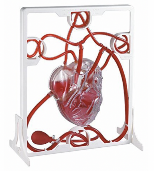
The Artsy® Human Heart Pumping Model transforms cardiovascular theory into visual, hands-on learning. Designed for educators, students, and medical professionals, this fully functional model simulates blood flow and heart contractions—bringing the circulatory system to life with every squeeze of the pump.
A Working Circulatory System You Can See and Feel
Unlike traditional static models, this dynamic tool replicates real-time blood movement through the heart’s chambers and major vessels. A manual pump simulates systole and diastole, circulating colored liquid through clear, anatomically aligned pathways, helping learners visualize the complete cycle of blood flow.
Key Features
Functional Pumping Simulation: Transparent heart chambers visibly contract to move liquid through the circulatory loop.
Color-Coded Circulation: Red and blue tubing distinguishes oxygenated and deoxygenated blood paths.
Manual Bulb Pump: Replicates the heartbeat, allowing full control over blood flow and rhythm.
Full Circulatory Circuit: Includes atria, ventricles, valves, arteries, and veins in one cohesive system.
Clear Acrylic Frame: Supports the model upright for optimal classroom visibility and stability.
What It Teaches
Use this model to demonstrate:
How blood flows through the heart and body
The difference between systemic and pulmonary circulation
The role of valves in preventing backflow
The direction of flow: vena cava → right atrium → right ventricle → lungs → left atrium → left ventricle → aorta
The effects of pressure changes or valve malfunction on circulation
Perfect For
Science Classrooms – Engage students in middle school through college with active learning.
Medical & Nursing Schools – Teach cardiac physiology, circulation, and valve function in real-time.
Clinics & Patient Education – Use visual aids to explain heart conditions clearly and memorably.
Homeschool & Lab Settings – Add tactile, STEM-focused tools to anatomy instruction.
Interactive & Kinesthetic Learning
The Artsy® Heart Pumping Model encourages participation:
Pump the bulb to simulate heartbeats
Watch liquid pass through each chamber
Trace circulation visually and spatially
Reinforce terminology and blood flow direction through movement-based memory
Ideal for kinesthetic and visual learners, this approach boosts comprehension and retention across STEM subjects.
Built to Teach and Last
Constructed with medical-grade plastic, flexible tubing, and an impact-resistant acrylic frame, this model is classroom-tough and designed for repeated hands-on use. It balances portability with visibility—perfect for both desktops and lecture demonstrations.
Specifications
Material: Medical-grade plastic and acrylic
Circulatory Tubing: Flexible, red/blue color-coded
Pump: Manual bulb-style
Display Size: Compact classroom size (varies slightly by batch)
Care: Wipe clean with cloth and mild disinfectant
Frequently Asked Questions
Does it actually pump liquid?
Yes. The bulb pump moves liquid visibly through the model’s transparent heart and vessel pathways.
Can the liquid be replaced?
Yes. The system is refillable with water-based, classroom-safe liquids.
Is it safe for younger students?
Yes—with supervision. It's durable and non-toxic, ideal for educational use at all levels.
Is this used in professional medical training?
Absolutely. It's widely adopted in medical and nursing education, EMT training, and patient instruction environments.
Practical Uses
Demonstrate systole/diastole and valve dynamics
Teach systemic vs. pulmonary circulation
Explain heart conditions like hypertension or valve disorders
Simulate how exercise affects heart rate and flow
Used By
High School & College Science Educators
Medical & Nursing Instructors
Cardiology Departments
Allied Health & EMT Trainers
Patient Education Centers
What Educators Say
“The students absolutely love it. Seeing the heart actually pump makes everything click.”
— Ms. Clarke, High School Biology Teacher
“I use it to explain valve disorders and flow resistance. Real-time visuals are game-changing.”
— Dr. Munir, Cardiology Educator
“Great for explaining heart failure in a way families can understand.”
— Nurse Practitioner, Cardiac Unit