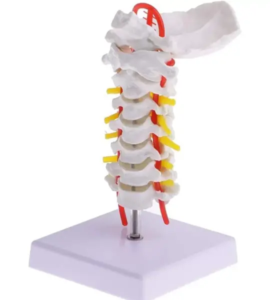
The cervical spine is a critical yet often misunderstood region of the human body. With its close proximity to the brainstem, its relationship with blood flow via vertebral arteries, and its role in head and neck movement, it deserves focused attention. The Cervical Vertebral Column With Neck Artery Model offers that clarity—through a compact, detailed, and easy-to-demonstrate replica of the upper spine.
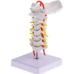
This anatomically accurate model of the cervical spine (C1 to C7) offers a clear view of each vertebra along with the path of the vertebral artery. It’s designed for clinical educators, students, and healthcare professionals who need a portable, high-detail cervical spine teaching aid.
Features:
Full cervical spine anatomy (C1–C7)
Highlighted vertebral artery route
Ideal for medical and chiropractic instruction
Compact, durable, and easy to transport
Suitable for classroom or clinical use
Applications:
✔ Clinical Teaching
✔ Patient Demonstration
✔ Medical Training
✔ Chiropractic Education
This life-like model features C1 to C7 vertebrae, complete with intervertebral discs, nerve roots, and the vertebral artery that runs through the transverse foramina. It includes the occipital bone at the top, providing a complete cervical-to-skull connection and making it a must-have for orthopedics, neurology, chiropractic training, physiotherapy education, and patient counseling.
Perfectly sized for desktops and OPD tables, the model rests on a clean white base and is mounted vertically, making it visible from all angles. Whether you're explaining a case of cervical spondylosis, herniation, or vascular compression, this tool adds tremendous value to your sessions.
“This cervical model helped me explain vertebral artery insufficiency to my students in a way a PowerPoint never could,” shares Dr. Kavita Sharma, Lecturer at GMC Aurangabad.
For students, the colored arteries and nerves serve as visual cues that make anatomical recall easier. For patients, it turns complex scans and symptoms into something they can understand at a glance. For educators, it brings practical anatomy to life—without needing a full spinal column model.
If you’re seeking a compact yet anatomically rich cervical spine model for academic or clinical use, MYASKRO’s offering stands tall—literally and figuratively—in any learning environment.
A Detailed View of the Cervical Spine – From C1 to Vertebral Artery
The Cervical Vertebral Column With Neck Artery Model offers a unique blend of compact size and high anatomical precision. Built specifically for focused cervical spine education, it includes critical elements like the occiput, vertebrae C1 through C7, intervertebral discs, and prominent visualizations of vertebral arteries and spinal nerves.
This model is ideal for demonstrating:
- Head and neck mobility linked to cervical structure
- The pathway of vertebral arteries through transverse foramina
- Nerve root exit patterns and potential impingement sites
- Postural dysfunction and cervical disc pathology
Each vertebra is molded with fine attention to detail—spinous processes, vertebral notches, and foramina are all realistically rendered. The arteries are highlighted in vibrant red while the spinal nerve roots are marked in yellow, making key structures instantly identifiable during explanation or instruction.
The compact footprint and upright mount make it easy to place on a table, podium, or consultation desk. Whether you’re running a lecture in a medical college or guiding a patient through symptoms of cervicogenic dizziness or radiculopathy, this model becomes a powerful point of reference.
Common applications include:
- Medical and physiotherapy classrooms for teaching cervical alignment
- Spine clinics to illustrate disc bulges or nerve compression
- Chiropractic setups for demonstrating vertebral artery involvement
The base is wide and non-slip, keeping the model stable during hands-on demonstrations. It’s made from durable, non-toxic PVC to withstand regular handling, whether by students, doctors, or instructors.
“We use it in every cervical spine lecture at Jamia Hamdard University. The vertebral artery depiction alone is worth it,” says Prof. A.R. Bhaskar, Neuroanatomy Department.
 Anatomy Models
 Ayurveda Models
 Charts
 Medical Simulators
 Women's Health Education
 OB/GYN Trainers
 Baby Care Simulators
 Medical/Emergency Rescue kit
 BAMS Lab Setup



.jpg)
