Khanna Traders, are producer and supplier of scientific equipment.
+91-8930598097
High-Precision Models
Education-Focused Designs
Wide Product Range
Trusted by Professionals
Long-lasting Material
Made in India Excellence
Have a question or need a custom anatomical model? Fill out the form below.
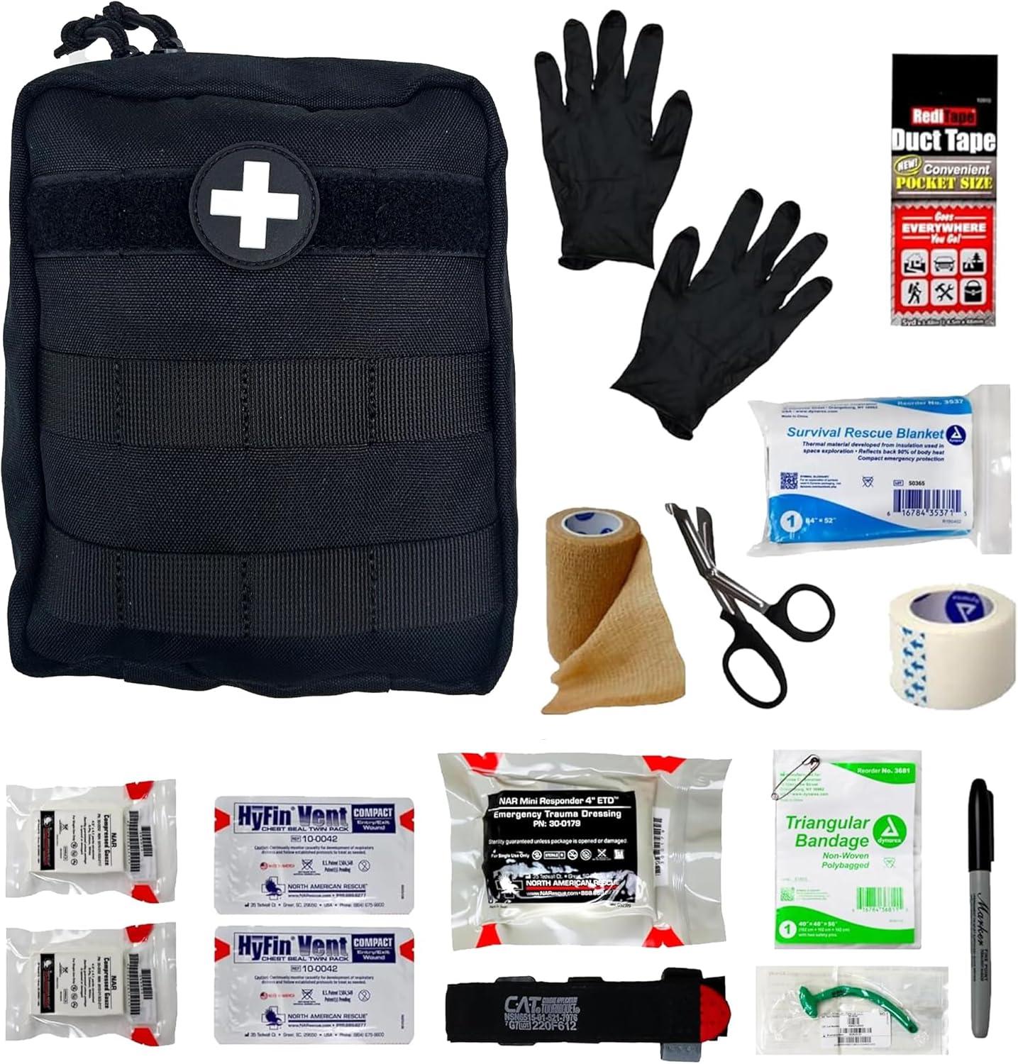
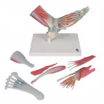
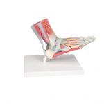
The foot skeleton features not only the bones but also the muscles, tendons, ligaments, nerves, arteries, and veins of the foot. The frontal view of the foot model features the extensor muscles of the lower leg. The tendons can be followed on their passage under the transverse and crucial crural ligaments all the way to their insertion points.
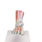
This anatomically detailed model of the foot and lower leg can be disassembled into 6 removable parts for detailed study of the foot and ankle. The foot skeleton features not only the bones but also the muscles, tendons, ligaments, nerves, arteries, and veins of the foot. The frontal view of the foot model features the extensor muscles of the lower leg. The tendons can be followed on their passage under the transverse and crucial crural ligaments all the way to their insertion points.
In addition all tendon sheaths of the foot area are visible. On the dorsal portion of the foot the gastrocnemius muscle is removable to reveal deeper anatomical elements. The sole of the foot is represented in three layers; the first layer displaying the flexor digitorum brevis. This muscle can be removed from the foot revealing the quadratus plantae, the tendon of the flexor digitorum longus, and the flexor hallucis muscle. This second layer is in turn removable to display even deeper anatomical details of the foot. This foot skeleton model with ligaments and muscles is the best of its kind in quality and value.
Your email address will not be published. Required fields are marked *
Please login to write review!
Looks like there are no reviews yet.
Understand how feet shape function. The Artsy Anatomical Foot Model Set features three life-size sagittal-section foot models: normal arch, high arch (pes cavus), and flat foot (pes planus). Each model is anatomically accurate, color-coded, and crafted for hands-on teaching and clinical clarity. Perfect for gait analysis, orthotic fitting, patient education, and anatomy instruction, this set helps explain how arch structure impacts posture, balance, and injury risk.
The Artsy Set of 3 Anatomical Foot Models makes one thing clear: the shape of the foot defines how the body moves.
From gait patterns to postural alignment, variations in arch height influence everything up the kinetic chain. This professional-grade model set includes three detailed, life-size cross-sectional foot replicas — normal, high-arched, and flat-footed — to support anatomy education, clinical diagnostics, and patient communication.
Each model showcases:
Bones: calcaneus, talus, navicular, metatarsals, phalanges
Ligaments: plantar fascia, deltoid ligament, and more
Tendons & Muscles: including the Achilles tendon
Sagittal View: interior structures and alignment clearly visible
Realistic Texture: color-coded relief and soft-touch durability
Normal Arch Model: Balanced tarsal alignment, optimal ligament tension, natural load distribution.
High Arch (Pes Cavus): Increased curvature, rigid structure, higher pressure zones — great for explaining stress fractures and plantar fasciitis.
Flat Foot (Pes Planus): Collapsed medial arch, overpronation, and ligament stretch — ideal for discussions on gait dysfunction and compensation injuries.
Whether you’re teaching biomechanics or prescribing orthotics, these side-by-side comparisons help students, patients, and professionals instantly recognize how foot structure affects overall function.
✅ Anatomical Accuracy – Developed with orthopedic and podiatric input
✅ Visual & Tactile Engagement – Designed to show and feel key structural differences
✅ Highly Durable Materials – Resists wear and fading even with daily use
✅ Portable and Desk-Friendly – Lightweight, stable, and travel-ready
✅ Ideal for Patient Education – Boosts understanding and treatment compliance
Medical & Allied Health Programs
Podiatric & Orthopedic Clinics
Sports Medicine & Gait Labs
Physical Therapy & Rehabilitation
Patient Consultations & Workshops
Biomechanics & Kinesiology Education
This set is already trusted by educators and professionals worldwide. Whether used during lectures, in OSCE prep, or as a visual aid in orthotic consultations, Artsy foot models transform complex biomechanical theory into clear, graspable insight.
“We use this set in every gait analysis session. It’s the fastest way to explain foot structure and its impact on movement.”
— Dr. Priya N., Sports Podiatrist, Melbourne
“Great visual tool. Clients understand their condition better, and compliance has gone up.”
— Amy B., Podiatrist & Clinic Owner, Toronto
This set naturally ranks for key educational and clinical search terms like:
flat foot anatomy model
high arch foot model
3D foot structure teaching tool
orthotic assessment training aid
plantar fascia visual model
Because that’s exactly what it is: a visual and tactile guide to foot mechanics in their most common forms — essential for anyone who teaches, diagnoses, or treats conditions tied to arch structure.
More than just models, this set is a dynamic teaching tool for real-world movement analysis. Show how an athlete’s high arch may increase impact stress. Explain how a flat foot contributes to knee pain. Demonstrate plantar fascia strain in seconds — no X-rays or charts required.
When the goal is lasting comprehension, the Artsy Anatomical Foot Model Set delivers. Quietly powerful, elegantly designed, and endlessly useful — it's an indispensable addition to any clinic or classroom.
The human spine is a marvel of alignment, articulation, and neural integration—and the Artsy 85 CM Vertebral Column Model brings this complexity to life with anatomical precision. Extending from the base of the skull to the tips of the femur heads, this life-size, full-length model is designed to teach, demonstrate, and visualize the entire spinal structure in an educational or clinical setting.
Featuring all 24 vertebrae—cervical, thoracic, and lumbar—plus the sacrum, coccyx, pelvis, and bilateral femur heads, this model includes critical anatomical markers such as:
A herniated lumbar disc
Clearly painted spinal nerve roots
The vertebral artery path
Intervertebral notches and foramina
At 85 cm in height, the model is mounted on a durable metal stand with a tripod base and top hook for easy 360° viewing. It’s an ideal asset for MBBS and BPT classrooms, physiotherapy rounds, OPD demonstrations, or chiropractic sessions.
“We introduced this model in our ortho OPD at ESIC Medical College, Hyderabad. It’s been incredibly useful in showing disc herniation and nerve root pressure to patients.” – Dr. Mansi Tripathi, Ortho Consultant
If you're preparing students for neuroanatomy exams, helping patients understand spinal issues, or training interns on postural mechanics, this model turns concepts into confident communication.
The Artsy Vertebral Column Model offers a high-resolution look at the entire axial skeleton, including:
Cervical vertebrae (C1–C7) – with atlas, axis, and odontoid process
Thoracic & lumbar vertebrae – spaced and shaped to reflect spinal curvature
Sacrum & coccyx – fused segments mounted securely to the pelvis
Pelvic girdle with femur heads – for demonstrating spinal load distribution
Additional features include:
Yellow spinal nerves emerging clearly between vertebrae
Herniated disc representation in the lumbar region
Vertebral artery passage through the cervical spine
Visible vertebral notches and intervertebral foramina
Whether you're teaching spinal curvature, diagnosing nerve impingement, or conducting hands-on practice, this model provides a comprehensive anatomical reference that enhances both learning and patient trust.
Physiotherapy & MBBS classrooms – full-spine orientation and palpation practice
Chiropractic & osteopathic clinics – structural demonstration for diagnosis
Orthopedic OPDs – patient-friendly tool to explain disc prolapse or sciatic pain
Neuroscience and radiology labs – understanding spinal cord and tract alignment
The reinforced PVC construction ensures durability during frequent handling, while the tripod metal base offers unmatched stability during presentations or exams.
“Our BPT batch at DY Patil University, Navi Mumbai found this model perfect for palpation practice and understanding spinal curvature patterns.” – Dr. Ritu Sinha, Lecturer, Department of Anatomy
The Artsy Vertebral Column Model is more than a teaching prop—it’s an interactive, practical tool that helps connect symptoms to spinal mechanics:
Cervical misalignment → nerve compression
Lumbar disc herniation → leg pain
Postural deviations → spinal loading patterns
By demonstrating these connections, educators and clinicians build stronger engagement with students and patients alike.
“We’ve been using this model daily in our neuro lab at AIIMS Raipur. It’s helped hundreds of students better understand spinal tract orientation and vertebral landmarks.” – Dr. Sameer Nanda, Professor of Anatomy
Life-size (85 cm) full spinal column with pelvis and femur heads
Includes herniated disc, nerve roots, arterial pathways, and vertebral notches
Tripod metal stand for stability and 360° visibility
Made from high-density, non-toxic PVC
Trusted by leading medical colleges, BPT programs, and OPDs across India
Whether you’re delivering a lecture on spinal curvature or guiding a patient through disc-related symptoms, the Artsy Vertebral Column With Pelvis Model gives you a clear, tactile, and durable resource for teaching and communication.
It’s not just a model—it’s a spinal education platform built for serious learning environments.
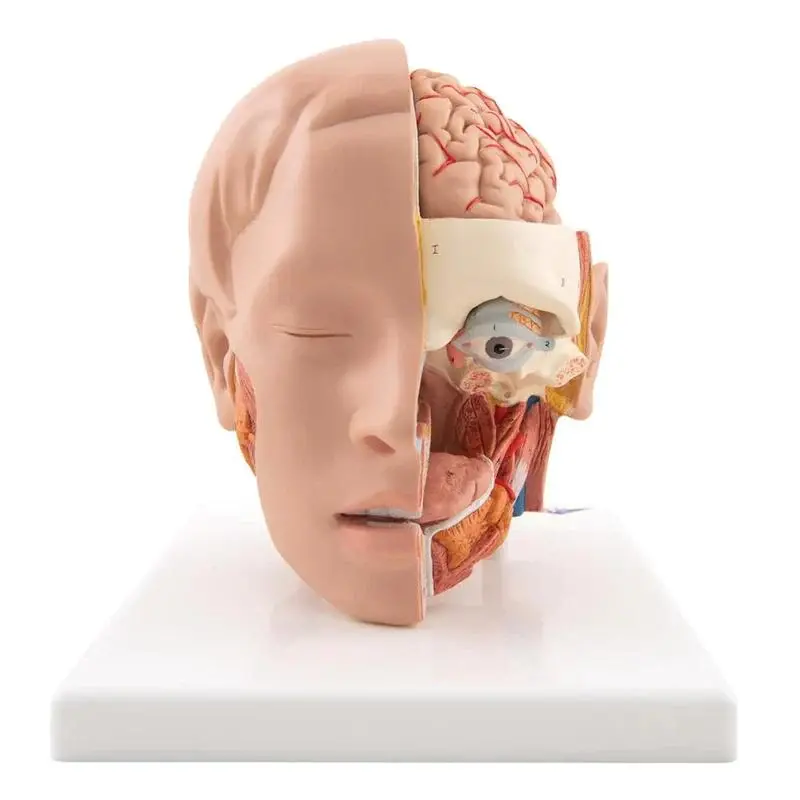
The Artsy Thoracic Spinal Column Model is a clinical-grade, full-scale replica of the thoracic spine, designed for precise anatomical education and effective patient communication. Covering vertebrae T1 through T12, it faithfully represents key thoracic structures including intervertebral discs, spinous and transverse processes, and bilateral yellow spinal nerve roots—offering a tangible, visual learning experience.
Whether used in a medical college classroom, a physiotherapy lab, or a spine care clinic, this model brings clarity to one of the most biomechanically complex yet commonly overlooked regions of the human body.
T1–T12 Vertebrae – Accurate size and curvature that reflect thoracic kyphosis
Spinous Processes – Angled downward, highlighting thoracic structural characteristics
Transverse Processes – Visible articulation points for demonstrating rib connections
Intervertebral Discs – Realistic spacing to simulate disc compression and height variation
Yellow Nerve Roots – Clearly painted to show bilateral nerve exit pathways
All elements are molded from high-density, non-toxic PVC, ensuring long-term durability with anatomical precision. The model is securely mounted on a sturdy base, allowing for hands-on demonstrations in academic and clinical settings.
Medical students preparing for anatomy practicals or OSCEs
Physiotherapy programs teaching thoracic biomechanics, posture, and mobility
Chiropractors and spine specialists explaining mid-back conditions to patients
Orthopedic and neurology educators demonstrating nerve impingement or structural misalignments
Teaching thoracic spinal structure and function
Demonstrating postural mechanics, thoracic nerve distribution, or disc-related conditions
Comparing with cervical and lumbar models to highlight vertebral differences
Patient education for kyphosis, scoliosis, costovertebral dysfunction, or nerve compression
Life-size proportions for clinical realism
High-visibility nerve paths help illustrate neuroanatomy with clarity
Lightweight yet durable—ideal for frequent handling in class or clinic
Compact enough for desk-side teaching, portable for mobile practitioners
Proven effective in both academic training and real-world diagnostics
“This model helped our BPT students at Manipal University grasp spinal nerve orientation and thoracic mobility limitations within minutes.” – Dr. Prerna Chhabra, Lecturer in Anatomy
“At our chiropractic campus in Pune, this model is used to teach everything from nerve root compression to vertebral body palpation.” – Prof. Ujjwal Mehta, Academic Coordinator
“Explaining thoracic pain patterns to patients used to take five minutes—now it takes thirty seconds with this model.” – Dr. Ananya Joshi, Spine Specialist, Apollo Clinic, Ahmedabad
This model aligns with top-searched educational and clinical terms like:
“Thoracic spine anatomy model”
“T1–T12 vertebrae teaching model”
“PVC thoracic vertebral column replica”
“Spinal nerve exit educational tool”
It’s already trusted by top medical colleges, physiotherapy schools, chiropractic centers, and orthopedic OPDs across India.
The Artsy Thoracic Spinal Column Model bridges the gap between theory and practical understanding. Whether you're walking students through spinal mechanics or guiding patients through treatment plans, this model enhances communication, reinforces accuracy, and promotes deeper anatomical insight.
It’s not just a demonstration tool—it’s an educational asset.
An Essential Tool for Teaching Lumbar Structure, Posture, and Pelvic Mechanics
The Artsy Pelvis with 5 Lumbar Vertebrae Model is a clinically accurate, life-size anatomical replica designed to bring the complexities of the lower spine and pelvic region into full focus. Whether you’re educating future healthcare professionals or guiding patients through treatment plans, this model provides a tangible, detailed reference for understanding lumbar structure, spinal nerves, and sacropelvic alignment.
Includes Five Lumbar Vertebrae (L1–L5)
Complete with realistic spinous processes, vertebral bodies, and transverse processes. Ideal for teaching lumbar anatomy, intervertebral spacing, and vertebral orientation.
Integrated Intervertebral Discs & Spinal Nerves
Clearly visible yellow nerves exit the spinal column at each vertebral level—perfect for demonstrating nerve root impingement and conditions like sciatica or spinal stenosis.
Full Pelvic Structure
Features the ilium, ischium, pubis, sacrum, and pubic symphysis in anatomical detail. Offers a visual reference for pelvic tilt, hip mechanics, and sacroiliac alignment.
Fixed Base for Stability
Mounted on a stable stand, this model is well-balanced for desk, table, or mobile display use in teaching, workshops, or clinical sessions.
Orthopedic, Chiropractic & Physiotherapy Education
Demonstrates spinal loading, pelvic stability, lumbar curve disorders (like lordosis), and neuromuscular relationships in 3D clarity.
OSCE and Practical Exam Preparation
Students can visually map vertebrae and nerve pathways—boosting retention and real-world understanding of lumbar neuroanatomy.
Patient Communication & Treatment Planning
Helps illustrate complex conditions like disc herniation, sacroiliac dysfunction, or lower back pain in a way that patients instantly grasp.
Constructed from Medical-Grade PVC
Durable, fade-resistant, and easy to clean—suitable for daily classroom or clinic use.
Anatomically Correct Scale
Life-size proportions ensure realistic spacing, joint relationships, and structure-to-structure visual comparisons.
Portable & Practical
Lightweight and compact enough for mobile instruction, yet detailed enough to remain a permanent reference in any lab or consultation room.
✔ Detailed spinal nerve exits for neuro-education
✔ Visual clarity in fast-paced teaching environments
✔ Trusted by physiotherapy schools, chiropractic colleges, and orthopedic educators worldwide
✔ A reliable demonstration aid for lumbar spine and pelvic alignment
“This model bridges theory and practice beautifully. I use it to teach both lumbar biomechanics and pelvic dysfunction—students learn faster, and patients understand better.”
— Dr. R. Kim, Osteopathic Instructor, Vancouver
“The clarity of vertebral spacing and the visible nerve roots make this model a standout in my anatomy lab. Durable, accurate, and accessible.”
— Dr. L. Menon, PT Educator, New Delhi
Pelvis and spine model with nerves
Lumbar vertebrae anatomical teaching aid
Spinal nerve impingement demonstration tool
Orthopedic lumbar model for patient education
Teaching lumbar anatomy shouldn’t require guesswork. With the Artsy Pelvis with 5 Lumbar Vertebrae, you gain a clear, reliable, and professional-grade model that elevates every lesson, consultation, or exam prep session. It’s not just a visual—it’s a hands-on learning experience.
Are you sure you want to remove this product from cart?