Khanna Traders, are producer and supplier of scientific equipment.
+91-8930598097
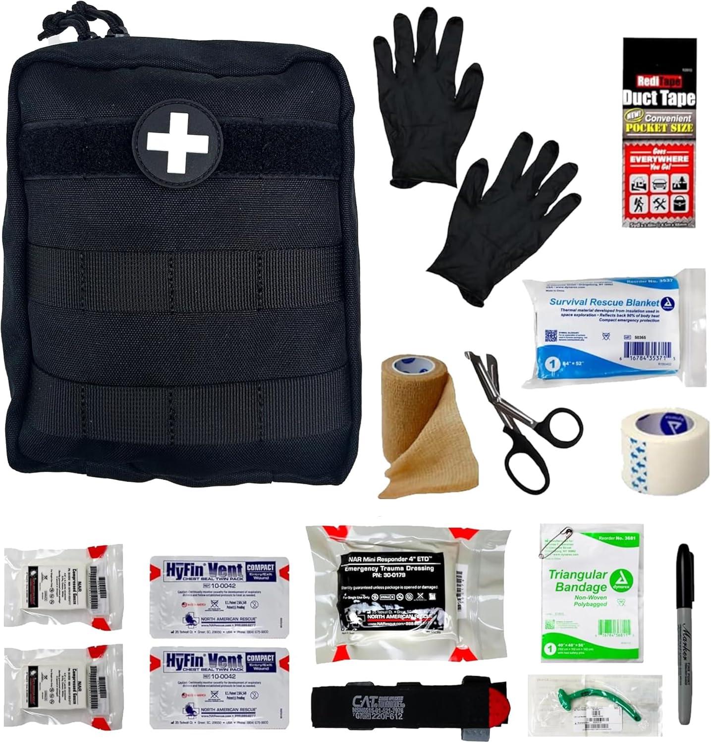
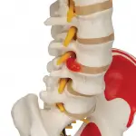
Understanding anatomy means seeing how systems interact—and the Human Skeleton Model with Nerves & Blood Vessels gives you that interaction in a compact, accessible format. With clearly marked arteries (red), veins (blue), and peripheral nerves (yellow) mapped across skeletal landmarks, this model offers a detailed multisystem overview that’s easy to move, easy to teach with, and endlessly valuable in both academic and clinical settings.
Standing at approximately 85cm tall (33 inches), this model is ideal for desktop teaching, one-on-one instruction, or visual reinforcement during clinical discussions. It includes major vascular routes and key neural paths that wrap around and pass through a fully articulated skeletal structure. From the carotid arteries and brachial plexus to the femoral nerve and iliac veins, each element is molded with clarity and anatomical precision.
Whether you're a medical student revising for practical exams, a biology teacher explaining human physiology, or a clinician helping patients understand their condition, this model delivers essential insights in a format that fits right on your desk.
Key benefits:
The Human Skeleton Model with Nerves & Blood Vessels goes beyond bone. It’s a compact, desk-friendly solution for teaching how the skeletal, circulatory, and nervous systems are structurally—and functionally—intertwined. While many models focus on just one element, this one integrates three, giving students and instructors a complete, real-world understanding of anatomy in action.
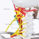
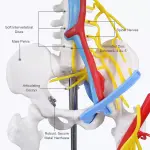
Each system is color-coded for clarity: bright red for arteries, blue for veins, and yellow for nerves. These structures are mapped along the skeleton in accurate anatomical pathways, helping learners trace critical routes like:
This layered design allows for cross-system instruction—such as explaining how a herniated disc may compress spinal nerves, or how the femoral triangle contains both vascular and neural elements crucial for clinical examination. It’s especially helpful in illustrating the impact of systemic conditions like stroke, peripheral artery disease, or nerve entrapment syndromes.
Though compact, this model includes impressive anatomical features:
In clinical environments, this model becomes an effective visual tool for patient education. Clinicians can point to arteries, nerves, and bones to explain diagnosis, treatment plans, or surgical procedures. Patients absorb complex information more quickly when they can see how structures interact, not just hear a description.
Its construction from unbreakable, washable PVC ensures durability and cleanliness, even with daily handling. Whether it’s being used in a bustling classroom or a consultation room, it holds up to the task—resistant to scratches, cracks, or staining.
As a nursing instructor shared, “This model changed how I teach anatomy. Students finally understand the relationships—because they can see them, clearly and all in one place.”
For educators and professionals alike, this is more than a skeleton. It’s a systematic learning platform built for simplicity, precision, and real-world insight.
Supplies and Replacement Parts:
Heavy Duty Dust Cover (W40103)
Metal Base With 5 Casters (XA031)
Your email address will not be published. Required fields are marked *
Please login to write review!
Looks like there are no reviews yet.