


Artsy Rodger Human Skull Model – Life-Size with Articulating Jaw & Removable Skull Cap
A Classic Yet Essential Tool for Medical Mastery
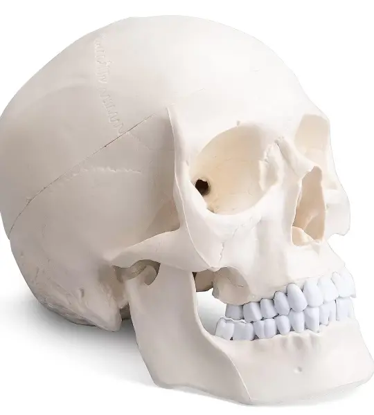
The Artsy Rodger Human Skull Model is a life-size, high-accuracy anatomical replica, crafted to support foundational learning in human cranial anatomy. Designed for medical, dental, and biology students, as well as for educators and healthcare professionals, this 3-part skull offers a hands-on learning experience with realistic structure and functionality.
Built with precision and care, this model includes a removable skull cap (calvaria) and an articulating mandible, providing full access to both external features and internal cranial anatomy.
🔍 Key Features
✅ Life-Size & Anatomically Accurate
Replicates the proportions and features of a real adult human skull, ideal for exam prep, viva practice, and demonstration.
✅ 3-Part Design for Enhanced Learning
Detachable Skull Cap: View internal cranial cavity and sinus structure
Articulating Jaw: Mandible is spring-held and movable, simulating natural jaw motion
Complete Dental Arch: Teeth are individually modeled, including incisors, molars, and canines
✅ High-Quality Material
Crafted from non-toxic, medical-grade PVC, durable enough for long-term use in classrooms or clinics.
✅ Ideal for Study & Display
Whether you're preparing for anatomy exams or decorating a doctor’s cabin, the Rodger Skull Model serves both academic and aesthetic purposes.
🧑🎓 Perfect For:
MBBS, BDS, Nursing & Paramedical Students
Anatomy Professors and Medical Institutions
Coaching Centers and Lab Demonstrators
Aspiring Doctors & Enthusiastic Learners
Gifting to Students Entering Medical Fields
📦 Specifications
Product Name: Artsy Rodger Human Skull Model
Design: 3-Part (Calvaria, Articulating Jaw, Base Skull)
Material: Premium Non-Toxic PVC
Scale: True 1:1 Life-Size
Weight: Approx. 1.1 kg
Finish: Natural matte texture for bone-like appearance
Inclusions: Skull model + Protective packaging
💬 What Our Users Say
“Simple, sturdy, and exactly what I needed for anatomy practicals.”
– Varsha P., BDS Student
“Looks great on my clinic desk—and even better when I need to explain cranial features to patients.”
– Dr. Anurag R., General Physician
🚚 Price & Delivery
MRP: ₹1,799 (Inclusive of Doorstep Delivery)
Return Policy: 3-Day Replacement Guarantee
Customer Support: 24x7 via support@artsyanatomy.com
Artsy – Learning That Lasts a Lifetime.
Whether it’s your first anatomy model or your fiftieth, the Rodger Skull delivers clarity, confidence, and clinical relevance.


