-
Khanna Traders, are producer and supplier of scientific equipment.
- khannachahat05@gmail.com
-
+91-8930598097
Khanna Traders, are producer and supplier of scientific equipment.
+91-8930598097
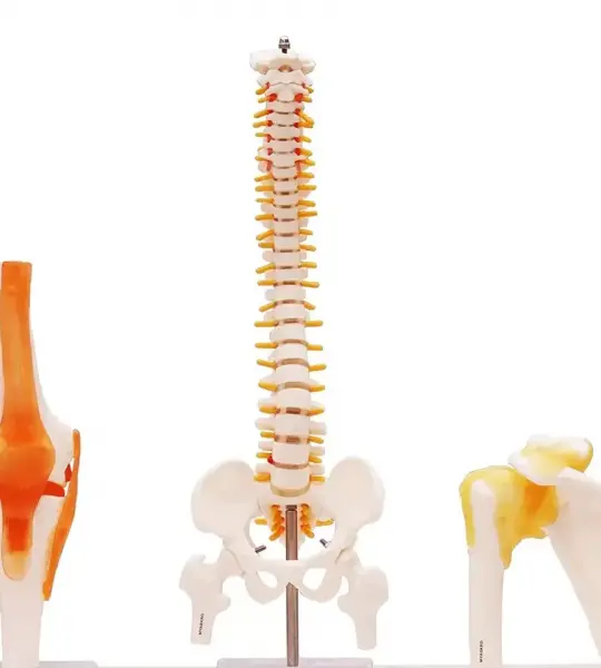
The Artsy Spine, Knee & Shoulder Anatomical Model Combo is a comprehensive educational toolkit designed for orthopedic instructors, medical students, physiotherapists, and clinical professionals. This trio covers the three most studied and movement-critical joints of the human body—spine, knee, and shoulder—offering up to 96% anatomical accuracy in a durable, portable format.
Spine Model: Showcases cervical to sacral vertebrae, spinal curvature, intervertebral discs, and nerve exits—ideal for teaching postural mechanics, neurological pathways, and spinal conditions.
Knee Model: Life-size and accurately labeled, it includes the femur, tibia, fibula, patella, and key ligaments such as the ACL and PCL. It’s perfect for explaining joint mechanics and common sports injuries.
Shoulder Model: Cast from an original specimen, this model displays the scapula, clavicle, humeral head, and associated tendons—enabling detailed instruction on rotator cuff injuries and arm movement.
Each model is mounted on a labeled stand, compact and easy to display on desks, tables, or treatment stations. They're fully assembled and ready for immediate use.
Educational Precision: Every model is crafted to replicate human anatomy with exceptional detail, allowing for real-time demonstration of motion, injury, and rehabilitation.
Clinical Relevance: Used by chiropractors, sports therapists, and rehabilitation specialists to enhance patient communication and understanding.
Durability: Made from medical-grade plastic, these models withstand frequent handling, making them suitable for daily use in both academic and clinical environments.
Time & Cost Savings: Purchasing the combo offers better value than buying models individually—and removes the hassle of sourcing separate pieces.
Orthopedic education and anatomy courses
Physiotherapy and rehabilitation training
Medical schools, biology classes, and clinical labs
Patient education in therapy and chiropractic settings
Educators: Praise its comprehensive coverage of essential joints used in weekly instruction.
Clinicians: Note its impact in explaining injuries and treatment plans effectively.
Students: Appreciate the tactile and visual clarity for mastering joint mechanics.
“These three models make up 80% of what I need for joint education. It’s a smart bundle—especially when you’re teaching motion and injury.”
Covers spinal alignment, knee stability, and shoulder mobility—the core of functional biomechanics.
Helps bridge the gap between theory and practice by allowing physical interaction and movement demonstration.
Offers clinical-level accuracy while remaining compact and accessible for classroom or bedside use.
Whether you're teaching OSCE prep, explaining rehab protocols, or demonstrating injury mechanics, the Artsy 3-in-1 Anatomical Set is your go-to toolkit for clear, confident instruction. It’s not just a set of models—it’s a mobile anatomy lab that reveals how the human body works in motion.
An Essential Tool for Teaching Lumbar Structure, Posture, and Pelvic Mechanics
The Artsy Pelvis with 5 Lumbar Vertebrae Model is a clinically accurate, life-size anatomical replica designed to bring the complexities of the lower spine and pelvic region into full focus. Whether you’re educating future healthcare professionals or guiding patients through treatment plans, this model provides a tangible, detailed reference for understanding lumbar structure, spinal nerves, and sacropelvic alignment.
Includes Five Lumbar Vertebrae (L1–L5)
Complete with realistic spinous processes, vertebral bodies, and transverse processes. Ideal for teaching lumbar anatomy, intervertebral spacing, and vertebral orientation.
Integrated Intervertebral Discs & Spinal Nerves
Clearly visible yellow nerves exit the spinal column at each vertebral level—perfect for demonstrating nerve root impingement and conditions like sciatica or spinal stenosis.
Full Pelvic Structure
Features the ilium, ischium, pubis, sacrum, and pubic symphysis in anatomical detail. Offers a visual reference for pelvic tilt, hip mechanics, and sacroiliac alignment.
Fixed Base for Stability
Mounted on a stable stand, this model is well-balanced for desk, table, or mobile display use in teaching, workshops, or clinical sessions.
Orthopedic, Chiropractic & Physiotherapy Education
Demonstrates spinal loading, pelvic stability, lumbar curve disorders (like lordosis), and neuromuscular relationships in 3D clarity.
OSCE and Practical Exam Preparation
Students can visually map vertebrae and nerve pathways—boosting retention and real-world understanding of lumbar neuroanatomy.
Patient Communication & Treatment Planning
Helps illustrate complex conditions like disc herniation, sacroiliac dysfunction, or lower back pain in a way that patients instantly grasp.
Constructed from Medical-Grade PVC
Durable, fade-resistant, and easy to clean—suitable for daily classroom or clinic use.
Anatomically Correct Scale
Life-size proportions ensure realistic spacing, joint relationships, and structure-to-structure visual comparisons.
Portable & Practical
Lightweight and compact enough for mobile instruction, yet detailed enough to remain a permanent reference in any lab or consultation room.
✔ Detailed spinal nerve exits for neuro-education
✔ Visual clarity in fast-paced teaching environments
✔ Trusted by physiotherapy schools, chiropractic colleges, and orthopedic educators worldwide
✔ A reliable demonstration aid for lumbar spine and pelvic alignment
“This model bridges theory and practice beautifully. I use it to teach both lumbar biomechanics and pelvic dysfunction—students learn faster, and patients understand better.”
— Dr. R. Kim, Osteopathic Instructor, Vancouver
“The clarity of vertebral spacing and the visible nerve roots make this model a standout in my anatomy lab. Durable, accurate, and accessible.”
— Dr. L. Menon, PT Educator, New Delhi
Pelvis and spine model with nerves
Lumbar vertebrae anatomical teaching aid
Spinal nerve impingement demonstration tool
Orthopedic lumbar model for patient education
Teaching lumbar anatomy shouldn’t require guesswork. With the Artsy Pelvis with 5 Lumbar Vertebrae, you gain a clear, reliable, and professional-grade model that elevates every lesson, consultation, or exam prep session. It’s not just a visual—it’s a hands-on learning experience.
A Complete, Clinical-Grade Representation of the Spinal Column
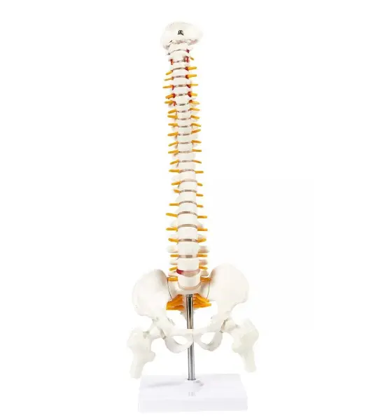
The Artsy Human Spine Model is an anatomically accurate, life-size teaching aid that incorporates the entire vertebral column from cervical to coccygeal regions, along with the pelvis and bilateral femur heads. Mounted on a robust stainless-steel stand, it offers educators, clinicians, and students a dynamic, tactile tool to explore spinal biomechanical function, pathology, and anatomical relationships.
Full-Length Anatomy
Includes all vertebrae (C1–C7, T1–T12, L1–L5), sacrum, coccyx, pelvis, and femur heads. Realistic spinal curves allow demonstrations of posture, alignment, and kinetic movement.
Herniated Disc Visualization
Features a prominently protruding lumbar disc to demonstrate nerve compression, disc pathology, and sciatica. It's a powerful visual asset for explaining clinical conditions.
Spinal Nerve Representation
Bright yellow spinal nerves exiting at each vertebral level offer clarity on neurological pathways and potential impingement.
Dynamic & Durable Design
Flexible column allows movement demonstration; supported by reinforced PVC and a sturdy chrome steel stand for durability in classroom and clinical settings.
Chiropractic & Physiotherapy Education
Illustrates spinal mechanics, postural curves (lordosis, kyphosis), and the impact of movement or misalignment.
Patient Consultation & Treatment Planning
Helps patients grasp the physical causes of back pain, disc herniation, and degenerative spine issues.
Medical & Allied Health Training
Supports neuroanatomy, orthopedic instruction, and hands-on exam preparation (MBBS, BPT, nursing, etc.).
Multi-Disciplinary Use
Ideal for orthopedists, neuro-spine specialists, fitness professionals, and massage therapists for workshops and demonstrations.
✔ Realistic and engaging: Students and patients interact more readily with a model they can manipulate.
✔ Clinically relevant: The herniated disc provides instant visual and tactile context for common pathologies.
✔ Portability meets stability: Despite its size, the model is durable and well-balanced for frequent use and movement.
✔ Semi-static yet dynamic: Emulates real-life spinal motion while retaining structural integrity.
Scale: Life-size, from skull base to coccyx
Construction: Reinforced PVC, high fidelity anatomical molding
Mounting: Stainless-steel stand with chrome finish, rotatable and stable
Intended Use: Teaching, clinical demonstrations, patient education
This is more than a decorative prop—it’s a functional learning platform. With precise vertebral detail, natural curvature, and clear nerve and disc depictions, the Artsy Spine Model enhances educational clarity, clinical insight, and professional communication. It empowers you to visually and physically map out spinal concerns, biomechanics, and therapeutic strategies—one vertebra at a time.
Visually Demystifying Bone Degeneration – Ideal for Education, Clinics & Research
Osteoporosis can be complex to explain—but the Artsy Cutaway Osteoporosis Model makes it clear. Featuring a cross-section of a bone (commonly femur or vertebra), this life-size model highlights the differences between healthy and osteoporotic bone structure. It’s meticulously designed to help medical professionals, physiotherapists, educators, and patients visually understand bone density loss and fracture risk.
✅ Realistic Bone Sections
Displays both healthy (dense, compact) and osteoporotic (porous, weakened) bone areas side-by-side
Enables direct comparison of trabecular (spongy) and cortical (outer shell) bone microstructures
✅ Cutaway Design for Enhanced Insight
Exposes cancellous bone interior—ideal for explaining micro-fracture susceptibility
Color-coded or textured layers may highlight advanced vs. early-stage osteoporosis
✅ Anatomical Accuracy
Modeled from actual bone scans or specimens
Ideal small-scale for easy review without needing full skeletal models
Clear representation of key anatomical landmarks and degeneration areas
✅ Durable, Safe & Portable
Made from high-grade, non-toxic PVC/resin
Lightweight and easy to carry—to classrooms, clinics, and workshops
Built to withstand frequent handling and patient demonstrations
Medical & Osteoporosis Clinics:
Communicate bone loss visually to help patients grasp diagnosis, fracture risk, and treatment importance
Physiotherapy & Rehab Centers:
Explain how weakened vertebrae or femoral necks respond to impact and load
Academic & Public Health Education:
Ideal for lectures, anatomy labs, awareness programs, and exam prep in MBBS, BPT, or nursing
Geriatric & Community Health Workshops:
Reinforce preventive messaging about nutrition and weight-bearing exercise
✔ Instant Visual Contrast: Seeing healthy vs. osteoporotic bone side-by-side fosters “aha” moments
✔ Supports Patient Compliance: Visual clarification boosts trust in management plans
✔ Exam-Ready Teaching Aid: Facilitates learning through tactile observation
✔ Compact & Classroom-Friendly: No need for large setups or separate high-tech tools
| Feature | Details |
|---|---|
| Model Type | Cutaway osteoporosis anatomy |
| Size & Scale | Life-size bone cross-section |
| Material | Medical-grade PVC or resin |
| Usage Environment | Clinics, classrooms, workshops |
| Portability | Lightweight & easy to transport |
The Artsy Cutaway Osteoporosis Anatomical Model bridges the gap between diagnosis and comprehension, transforming abstract scans and jargon into a clear, visible learning experience. Perfect for patient education, student teaching, and public awareness, it’s a small model with a significant educational impact.
Where Anatomy Meets Clarity – Teach, Demonstrate, and Empower with Confidence
The Artsy Lumbar Vertebrae with Sacrum, Coccyx & Herniated Disc Model brings the intricacies of the human lower spine to life. Designed for orthopedic professionals, physiotherapists, chiropractors, anatomy educators, and students, this life-size model offers a remarkably detailed and tactile view of the lumbosacral region.
Featuring five lumbar vertebrae (L1–L5), intervertebral discs, sacrum, coccyx, and realistic yellow spinal nerves, it provides a comprehensive anatomical overview. A standout feature is the raised, vividly colored herniated disc at L4-L5, giving users a powerful tool to explain disc pathology, nerve impingement, and sciatic pain with unmatched visual impact.
Whether used in anatomy classrooms, rehab centers, consultation rooms, or mobile clinics, this model is compact yet rich in detail—transforming spinal education from theory into tangible understanding.
🦴 Complete Lumbosacral Segment
Includes L1–L5 vertebrae, intervertebral discs, sacrum, and coccyx
Ideal for showing spinal curvature, nerve pathways, and pelvic transition
Supported by a stable steel-mounted base for 360° demonstration
📛 Realistic Herniated Disc at L4–L5
Textured, red-highlighted protrusion simulates disc bulge with accuracy
Clearly shows how herniation affects adjacent spinal nerve roots
Perfect for explaining common issues like sciatica and lower back pain
🧠 Educational Utility Across Disciplines
For orthopedic consultations, chiropractic sessions, and physiotherapy evaluations
Excellent teaching tool for BPT, MBBS, and allied health anatomy curricula
Students can palpate vertebral structures, understand spinal mechanics, and memorize landmarks efficiently
🧳 Compact Yet Clinically Insightful
Lightweight and easy to carry for workshops, mobile demos, or campus rounds
Designed for daily clinical or academic use—resistant to stains, chips, and deformation
PVC-based synthetic resin ensures lasting quality and safety
Orthopedic and neuro consultations – visually explain lumbar disc issues
Rehabilitation and pain clinics – support patient compliance through clarity
Medical and nursing education – offer students a 3D tactile learning experience
Postural education sessions – reinforce understanding of spinal support and flexibility
“It’s helped me explain disc issues to patients far more effectively. They understand it the moment they see it.”
— Dr. Kamal V., Orthopedic Consultant, Delhi
✔ Life-size and anatomically precise
✔ Clearly marked spinal nerves and foramina
✔ Visualizes the transition from spine to pelvis
✔ Easy to transport, easy to understand, hard to forget
The Artsy Lumbar Vertebrae with Herniated Disc Model isn’t just an anatomical prop—it’s a daily-use asset that improves communication, learning, and decision-making. Its compact size fits any space, but its educational power is immense. When explaining spinal compression, disc degeneration, or nerve root impingement, no diagram or scan compares to the hands-on clarity this model offers.
From orthopedic surgeons and physiotherapists to anatomy professors and patient educators, this tool is trusted by professionals who value simplicity, accuracy, and impact.
“It’s one of the most used models in our training center. Explaining back pain, posture, or disc injuries becomes far easier with this on hand.”
— Dr. Lee M., Clinical Educator, Singapore
Model Includes: L1–L5 vertebrae, herniated disc, sacrum, coccyx, spinal nerves
Material: Medical-grade, non-toxic PVC
Mounting: Fixed on a stable steel rod and base
Size: Life-size dimensions for anatomical accuracy
Weight: Lightweight and portable
Artsy's life-size lumbar spine model demonstrates more than just anatomy—it builds trust, clarifies complexity, and improves outcomes. Whether you're guiding a patient through their condition or helping a student prepare for exams, this model delivers learning that lasts.
Explore the Core of Spinal Mechanics – Realistic, Robust, and Ready to Teach
The Artsy Lumbar Vertebrae Model (4pcs) offers a high-fidelity, life-size representation of the lower spine’s structural anatomy. Designed for medical educators, physiotherapists, chiropractors, and orthopedic professionals, this model provides a focused, hands-on way to understand the lumbar region—one of the most clinically significant areas of the vertebral column.
With four individual vertebrae from the lumbar spine (typically L1–L4), this model allows for in-depth demonstrations of spinal curvature, vertebral alignment, disc spacing, and load-bearing mechanics. It’s a must-have tool for teaching students and explaining conditions such as herniation, stenosis, or spondylolisthesis to patients.
🦴 4 Life-Size Lumbar Vertebrae
Includes four separate vertebrae (modeled after L1 to L4)
Precise anatomical landmarks: vertebral bodies, spinous and transverse processes, pedicles, laminae, and superior/inferior articular facets
Allows comparative study between segments for pattern recognition and clinical correlation
📐 True-to-Life Proportions
Cast from real human vertebrae specimens
Demonstrates correct curvature and size relative to actual human anatomy
Supports palpation training and hands-on examination
📚 Multi-Disciplinary Teaching Tool
Ideal for orthopedics, chiropractic studies, physical therapy, and neuroanatomy education
Helps visualize mechanical stress points and understand lumbar pathologies
Supports education in posture-related dysfunction, disc degeneration, and nerve impingement
🧳 Durable, Portable & Classroom-Ready
Made from premium-grade, non-toxic PVC plastic
Lightweight and easy to transport—perfect for field teaching, clinics, or lab work
Durable for repeated use in high-traffic educational settings
Anatomy & Physiology Courses – For MBBS, BPT, Nursing, and Allied Health programs
Clinical Counseling – To explain lumbar pain, disc problems, and spinal treatments to patients
Practical Exams & Viva Prep – Students can identify landmarks and practice anatomical reasoning
Chiropractic & PT Training – Use during spinal assessment and manual therapy demonstrations
“The 4-piece lumbar model from Artsy is one of our most-used tools in musculoskeletal lab sessions. Its accuracy and durability make it ideal for daily student handling.”
— Prof. Vikram Solanki, Department of Physiotherapy, AIIMS Jodhpur
✔ Life-size, cast from real human specimens
✔ Supports multiple teaching styles—visual, tactile, and kinesthetic
✔ Clearly defined bony landmarks for exam prep and clinical education
✔ Compact and easy to handle—no setup required
Whether you're explaining the biomechanics of lifting injuries, teaching disc alignment, or preparing students for clinical rotations, the Artsy Lumbar Vertebrae (4pcs) Life-Size Model offers the anatomical realism and durability your work demands.
Focused, Functional, Foundational – A Trio of Lumbar Vertebrae for Targeted Teaching
The Artsy Lumbar Vertebrae (3pcs) Anatomical Model Set is a compact, high-precision educational tool designed to help medical professionals, educators, and students understand the unique structural features of the lumbar spine. Each vertebra in this three-piece set is individually molded and anatomically accurate, showcasing the robust, weight-bearing characteristics of the L1–L5 spinal region.
This model is perfect for institutions, physiotherapy programs, chiropractic demonstrations, and orthopedic clinics where a focused examination of the lumbar vertebrae is essential.
🦴 Anatomically Accurate Lumbar Vertebrae
Includes three separate lumbar vertebrae (L-type)
Captures key features such as thick vertebral bodies, short spinous processes, and large vertebral foramina
Ideal for teaching spinal loading, lumbar lordosis, and mechanical function
🔍 Detailed Bony Landmarks
Clearly visible transverse processes, pedicles, laminae, and articular facets
Accurate texture and contour for tactile learning
Helps students identify vertebral features quickly in exams and lab sessions
🧠 Designed for Multi-Disciplinary Use
Useful in anatomy, orthopedics, neurology, and physiotherapy training
Supports clinical demonstrations of spinal loading, lower back pain mechanics, and vertebral degeneration
Allows comparison between normal and pathological alignment (if paired with other models)
📏 Compact, Durable & Classroom-Friendly
Made from high-quality, non-toxic PVC for durability
Easy to handle, pass around in classrooms, or use in hands-on practical exams
Lightweight, portable design – perfect for teaching on the go
Medical Education – For MBBS, BPT, and nursing programs learning spinal anatomy
Chiropractic Training – To demonstrate lumbar segment function and manipulation
Clinical Demonstration – Used by spine specialists for patient education about lumbar issues
Museum & Lab Display – Adds depth to anatomical collections in teaching environments
“The Artsy 3-piece lumbar set helps our students understand vertebral variations and spatial orientation faster than flat diagrams ever could.”
— Dr. Sujata Nair, Anatomy Professor, DY Patil Medical College
✔ Realistic structure and texture for hands-on learning
✔ Ideal size and portability for classroom and clinical use
✔ Supports tactile, visual, and kinesthetic learning styles
✔ A versatile, cost-effective addition to any spine model collection
Focus on the Foundation of the Spine – Teach Lumbar Anatomy with Clarity and Confidence
Whether you're breaking down the biomechanics of lumbar flexion or highlighting the structural basis of lower back pain, the Artsy Lumbar Vertebrae (3pcs) Anatomical Model Set gives you the clarity, accuracy, and portability you need. Built to last and designed to teach.
Transforming Spinal Education Through Visual Clarity and Hands-On Learning
The Artsy Lumbar Spine Herniated Disc Prolapse Model is a compact anatomical teaching aid designed to vividly illustrate one of the most common spinal pathologies—lumbar disc herniation. With lifelike vertebrae, intervertebral discs, and clearly visible nerve roots, this model offers an intuitive and tactile explanation of nerve compression, disc bulging, and lower back pain.
Compact enough to fit in the palm of your hand, yet detailed enough for academic and clinical accuracy, this model is an essential tool for orthopedic educators, neurospine specialists, physiotherapists, and students of anatomy.
🔴 Realistic Disc Herniation Simulation
Includes two lumbar vertebrae with a vivid, red bulging disc to depict prolapse
Clearly shows how the herniated disc impinges on adjacent spinal nerves
Designed for easy demonstration of sciatica, nerve compression, and back pain mechanisms
🟡 Detailed Nerve Anatomy
Spinal nerve roots emerge from the foramina with accurate color and structure
Helps explain radiating pain, numbness, and neurological symptoms
Ideal for neurology OPDs, spine consultations, and neuroanatomy courses
🔵 True-to-Form Vertebrae Construction
Anatomically shaped vertebrae with pedicles and spinous processes
Intervertebral space realistically represented for demonstrating compression
Clearly defined spinal canal and foramina for enhanced visibility
🟢 Built for Versatility and Portability
Made of medical-grade, non-toxic PVC
Lightweight (approx. 1.2kg) and palm-sized (6.7 x 5.1 x 6.7 inches)
Fits easily in a medical bag, classroom kit, or OPD counter
Orthopedic & Neurosurgical Clinics – For pre-op explanations and patient counselling
Physiotherapy Centers – To demonstrate movement restrictions and rehabilitation planning
Medical & Paramedical Education – In anatomy labs, lectures, and viva preparations
Public Health Settings – At awareness camps, physiotherapy workshops, or community events
“Using the Artsy herniation model during OPD sessions saves time and improves patient understanding. It’s much easier than explaining MRI reports,” — Dr. Ishaan Kumar, Neurospine Consultant, PGI Chandigarh
✅ Bridges the Gap Between Imaging and Understanding
Patients often struggle to connect radiological findings to symptoms. This model makes it simple, immediate, and relatable.
✅ Shortens the Learning Curve
Students preparing for orthopedic or neuro modules grasp spinal pathology more quickly with a hands-on, 3D representation.
✅ Supports Better Patient Compliance
When patients visually see the pinched nerve and bulging disc, their trust in the recommended treatment increases.
✅ Durable and Classroom-Tested
Crafted for frequent use, this model maintains its structural integrity over time—ideal for daily use in clinics and institutions.
The Artsy Lumbar Herniated Disc Prolapse Model is more than a visual aid—it’s a teaching partner. It demystifies complex lumbar spine conditions, improves communication between practitioner and patient, and enhances classroom learning with a tangible, clear, and engaging experience.
Whether you're explaining lumbar nerve compression to a patient or preparing a student for their anatomy practical, this model provides the accuracy and visual impact needed to make spinal education simple and memorable.
Precision-Crafted for Clarity, Accuracy, and Clinical Relevance
The Artsy Human Spine Model is a compact, 45cm anatomical replica designed for maximum educational value. Featuring lifelike vertebrae, femur heads, spinal nerves, a pelvis, and a herniated lumbar disc, this model offers exceptional detail and anatomical accuracy for educators, students, chiropractors, and clinicians.
Crafted under the supervision of experienced anatomists, the Artsy spine model delivers 96% anatomical accuracy and is molded directly from real human vertebrae specimens. It’s a trusted tool for teaching spinal structure, common injuries, postural issues, and nerve-related disorders.
🔹 Anatomically Accurate Design
Developed with input from expert anatomists
Precision-molded from real human spine specimens
96% alignment with actual human vertebral anatomy
🔹 Complete Educational Detail
Includes spinal nerves for neurology education
Features a herniated lumbar disc for clinical demonstration
Pelvis and femur heads included to show hip-spine interaction
True-to-life curves and intervertebral spacing
🔹 Tailored for Clinical Educators
Designed in consultation with chiropractors and spine care professionals
Ideal for explaining spinal dysfunctions like disc bulges or nerve compression
Perfect for chiropractic adjustments, orthopedic teaching, and physical therapy instruction
🔹 Durable, High-Quality Build
Made from non-toxic, medical-grade PVC
Lightweight (1.2 kg) and portable for classroom, clinic, or seminar use
Stable construction ensures longevity through repeated handling
Product Name: Artsy Human Spine Model
Height: 45 cm
Material: Non-Toxic PVC
Weight: 1.2 kg
Components: Spine, pelvis, femur heads, spinal nerves, herniated lumbar disc
Intended Use: Medical teaching, clinical demonstration, student education, patient explanation
Chiropractic Clinics – Demonstrate misalignment, disc herniation, and treatment outcomes
Medical Education – Teach vertebral anatomy, spinal pathology, and nerve distribution
Physiotherapy Training – Explain biomechanics and posture-related dysfunctions
Patient Counseling – Enhance visual understanding of diagnosis and therapy plans
“This is one of the most detailed compact spine models we’ve used in our clinic. The inclusion of nerves and herniated disc makes it far more useful for real-world explanation.”
— Dr. R. Iyer, Spine & Sports Rehab Specialist, Mumbai
Whether you’re teaching spinal mechanics to students or helping a patient understand a nerve impingement, the Artsy Human Spine Model provides clarity, credibility, and confidence. Designed with educators, doctors, and chiropractors in mind, it’s the compact yet powerful tool for spinal education.
Transform the Way You Teach and Understand the Spine
The Artsy Human Spine Didactic Anatomical Model offers an unmatched blend of anatomical accuracy, color-coded clarity, and durable construction—ideal for medical teaching, clinical demonstration, and hands-on learning. Life-sized and crafted from medical-grade materials, this model brings spine anatomy to life by clearly distinguishing each spinal region with vibrant, purpose-driven colors.
Color-Coded for Clarity
Each vertebral region is distinctly painted to support rapid identification and functional understanding:
Orange: Cervical Spine (C1–C7)
Blue: Thoracic Spine (T1–T12)
Red: Lumbar Spine (L1–L5)
Yellow: Sacrum & Coccyx
Anatomical Features:
24 vertebrae with accurate curvature and spacing
Intervertebral discs between each segment
Fused sacrum and coccyx
Paired femoral heads for pelvic orientation
Nerve roots exiting laterally to show clinical pathways
Mounted on a stable stainless steel tripod stand for full 360° visibility
Functional Design for Real-World Teaching
This is more than a static spine replica. Each vertebra is individually molded and hand-painted to ensure anatomical precision. The model supports dynamic instruction—from explaining scoliosis or lordosis to demonstrating disc bulges or nerve compression syndromes. With full lumbopelvic anatomy included, it enables detailed discussions around core stability, postural imbalances, and spinal alignment.
Versatile Educational Applications:
Medical colleges & coaching centers – for anatomy labs and viva prep
Physiotherapy & chiropractic clinics – for patient education and movement analysis
Orthopedic & neuro-spine consultations – for pre-op and rehab explanations
Fitness & rehabilitation programs – for teaching spine-safe mechanics
Why Educators Choose Artsy:
Durable for repeated classroom and clinical use
Professional color coding enhances student retention
Realistic nerve and disc anatomy aid clinical relevance
Sturdy, portable, and easy to reposition during sessions
What Professionals Say:
“We use this in biomechanics classes at LNCT Bhopal. The color coding makes viva prep faster and far more intuitive.” – Prof. Sharanya Mehta, PT Faculty
“At Manipal Spine Centre, it helps patients visualize back pain causes in a way X-rays can’t. It's become essential to our counselling sessions.” – Dr. Amit Kelkar, Neurospine Consultant
Whether you’re guiding a medical student through neuroanatomy or explaining sciatica to a patient, the Artsy Didactic Spine Model offers a clear, accurate, and memorable experience. Designed to simplify complexity, this model is your trusted visual tool—bridging the gap between theory and real-world understanding.
Built to Teach. Designed to Last. Trusted by Experts.
Designed for medical students, physiotherapists, orthopedic specialists, chiropractors, and fitness educators, this model allows learners to examine how vertebrae align, how muscles act on bone, and how flexibility, posture, and nerve pathways all work together in the spine’s operation. The entire spinal column—from cervical to coccygeal—is represented with life-like curvature and movement, and the model flexes naturally to replicate bending, twisting, and real-world spinal motion.
Key features include:
For student labs, the model reinforces textbook learning by giving learners a tangible, mobile frame to manipulate and explore. For clinical professionals, it becomes an indispensable patient education tool—illustrating the mechanics of back pain, disc herniation, postural misalignment, and muscular strain with real-time movement and reference points.
Mounted securely on a supportive stand, the model remains upright for display but detaches easily for hands-on demonstrations. Its durable materials and anatomical fidelity make it suitable for daily handling, in-depth instruction, and detailed biomechanical discussion.
As one chiropractor shared, “This model lets me show patients what their spine is doing—and why it matters. It turns anatomy into action.”
Spinal Movement, Muscular Function, and Nerve Pathways—All in One Model
The Flexible Spine Model delivers the trifecta of functional anatomy—skeletal structure, muscular attachment, and neural integration—all with interactive realism. This model is engineered not only to demonstrate static anatomy, but to give learners and practitioners a clear, hands-on understanding of how the spine moves, how muscles pull, and how nerves flow through it all.
What sets this model apart is its full flexibility. You can bend, rotate, and manipulate the spine to show cervical extension, thoracic kyphosis, lumbar lordosis, or sacral curvature. This flexibility is particularly useful when teaching spinal mechanics, postural correction, or injury patterns such as disc herniation or nerve impingement.
Every detail is carefully designed for educational depth:
This model becomes especially powerful in environments where active demonstration is key:
The flexibility also allows educators to reinforce concepts like spinal segment mobility, intervertebral disc function, and the effects of movement restriction. By observing how the model flexes or resists motion, learners build intuitive understanding—not just memorized terms.
Constructed from high-quality, medical-grade materials, the model is built for years of repeated use. The flexible column holds shape while allowing smooth articulation, and the colored markers stay vivid after countless demonstrations.
As one physiotherapy lecturer said, “It’s not just a model—it’s a conversation starter. Students suddenly ‘see’ what the spine is doing beneath the skin.”
Flexible Spine Model offers a dynamic, realistic way to teach spinal anatomy and function. Unlike rigid models, it demonstrates natural movement—bending, rotating, and compressing—alongside clearly marked muscle origins (red) and insertions (blue), visible nerve pathways, and removable femur heads for full lumbopelvic education.
Key Benefits:
Ideal for:
Orthopedic educators, chiropractors, physical therapists, fitness professionals, and anyone teaching spinal mechanics or rehabilitation.
Why It Stands Out:
Transforms static memorization into dynamic understanding—bringing anatomy, movement, and clinical relevance together in one essential teaching tool.
The cervical spine is a critical yet often misunderstood region of the human body. With its close proximity to the brainstem, its relationship with blood flow via vertebral arteries, and its role in head and neck movement, it deserves focused attention. The Cervical Vertebral Column With Neck Artery Model offers that clarity—through a compact, detailed, and easy-to-demonstrate replica of the upper spine.
This anatomically accurate model of the cervical spine (C1 to C7) offers a clear view of each vertebra along with the path of the vertebral artery. It’s designed for clinical educators, students, and healthcare professionals who need a portable, high-detail cervical spine teaching aid.
Features:
Full cervical spine anatomy (C1–C7)
Highlighted vertebral artery route
Ideal for medical and chiropractic instruction
Compact, durable, and easy to transport
Suitable for classroom or clinical use
Applications:
✔ Clinical Teaching
✔ Patient Demonstration
✔ Medical Training
✔ Chiropractic Education
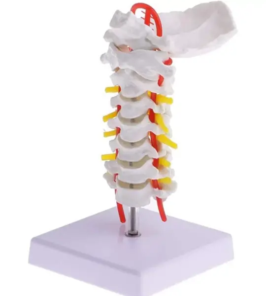
This life-like model features C1 to C7 vertebrae, complete with intervertebral discs, nerve roots, and the vertebral artery that runs through the transverse foramina. It includes the occipital bone at the top, providing a complete cervical-to-skull connection and making it a must-have for orthopedics, neurology, chiropractic training, physiotherapy education, and patient counseling.
Perfectly sized for desktops and OPD tables, the model rests on a clean white base and is mounted vertically, making it visible from all angles. Whether you're explaining a case of cervical spondylosis, herniation, or vascular compression, this tool adds tremendous value to your sessions.
“This cervical model helped me explain vertebral artery insufficiency to my students in a way a PowerPoint never could,” shares Dr. Kavita Sharma, Lecturer at GMC Aurangabad.
For students, the colored arteries and nerves serve as visual cues that make anatomical recall easier. For patients, it turns complex scans and symptoms into something they can understand at a glance. For educators, it brings practical anatomy to life—without needing a full spinal column model.
If you’re seeking a compact yet anatomically rich cervical spine model for academic or clinical use, MYASKRO’s offering stands tall—literally and figuratively—in any learning environment.
A Detailed View of the Cervical Spine – From C1 to Vertebral Artery
The Cervical Vertebral Column With Neck Artery Model offers a unique blend of compact size and high anatomical precision. Built specifically for focused cervical spine education, it includes critical elements like the occiput, vertebrae C1 through C7, intervertebral discs, and prominent visualizations of vertebral arteries and spinal nerves.
This model is ideal for demonstrating:
Each vertebra is molded with fine attention to detail—spinous processes, vertebral notches, and foramina are all realistically rendered. The arteries are highlighted in vibrant red while the spinal nerve roots are marked in yellow, making key structures instantly identifiable during explanation or instruction.
The compact footprint and upright mount make it easy to place on a table, podium, or consultation desk. Whether you’re running a lecture in a medical college or guiding a patient through symptoms of cervicogenic dizziness or radiculopathy, this model becomes a powerful point of reference.
Common applications include:
The base is wide and non-slip, keeping the model stable during hands-on demonstrations. It’s made from durable, non-toxic PVC to withstand regular handling, whether by students, doctors, or instructors.
“We use it in every cervical spine lecture at Jamia Hamdard University. The vertebral artery depiction alone is worth it,” says Prof. A.R. Bhaskar, Neuroanatomy Department.