-
Khanna Traders, are producer and supplier of scientific equipment.
- khannachahat05@gmail.com
-
+91-8930598097
Khanna Traders, are producer and supplier of scientific equipment.
+91-8930598097
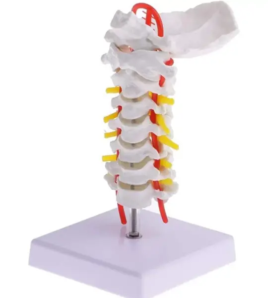
The cervical spine is a critical yet often misunderstood region of the human body. With its close proximity to the brainstem, its relationship with blood flow via vertebral arteries, and its role in head and neck movement, it deserves focused attention. The Cervical Vertebral Column With Neck Artery Model offers that clarity—through a compact, detailed, and easy-to-demonstrate replica of the upper spine.
This anatomically accurate model of the cervical spine (C1 to C7) offers a clear view of each vertebra along with the path of the vertebral artery. It’s designed for clinical educators, students, and healthcare professionals who need a portable, high-detail cervical spine teaching aid.
Features:
Full cervical spine anatomy (C1–C7)
Highlighted vertebral artery route
Ideal for medical and chiropractic instruction
Compact, durable, and easy to transport
Suitable for classroom or clinical use
Applications:
✔ Clinical Teaching
✔ Patient Demonstration
✔ Medical Training
✔ Chiropractic Education
This life-like model features C1 to C7 vertebrae, complete with intervertebral discs, nerve roots, and the vertebral artery that runs through the transverse foramina. It includes the occipital bone at the top, providing a complete cervical-to-skull connection and making it a must-have for orthopedics, neurology, chiropractic training, physiotherapy education, and patient counseling.
Perfectly sized for desktops and OPD tables, the model rests on a clean white base and is mounted vertically, making it visible from all angles. Whether you're explaining a case of cervical spondylosis, herniation, or vascular compression, this tool adds tremendous value to your sessions.
“This cervical model helped me explain vertebral artery insufficiency to my students in a way a PowerPoint never could,” shares Dr. Kavita Sharma, Lecturer at GMC Aurangabad.
For students, the colored arteries and nerves serve as visual cues that make anatomical recall easier. For patients, it turns complex scans and symptoms into something they can understand at a glance. For educators, it brings practical anatomy to life—without needing a full spinal column model.
If you’re seeking a compact yet anatomically rich cervical spine model for academic or clinical use, MYASKRO’s offering stands tall—literally and figuratively—in any learning environment.
A Detailed View of the Cervical Spine – From C1 to Vertebral Artery
The Cervical Vertebral Column With Neck Artery Model offers a unique blend of compact size and high anatomical precision. Built specifically for focused cervical spine education, it includes critical elements like the occiput, vertebrae C1 through C7, intervertebral discs, and prominent visualizations of vertebral arteries and spinal nerves.
This model is ideal for demonstrating:
Each vertebra is molded with fine attention to detail—spinous processes, vertebral notches, and foramina are all realistically rendered. The arteries are highlighted in vibrant red while the spinal nerve roots are marked in yellow, making key structures instantly identifiable during explanation or instruction.
The compact footprint and upright mount make it easy to place on a table, podium, or consultation desk. Whether you’re running a lecture in a medical college or guiding a patient through symptoms of cervicogenic dizziness or radiculopathy, this model becomes a powerful point of reference.
Common applications include:
The base is wide and non-slip, keeping the model stable during hands-on demonstrations. It’s made from durable, non-toxic PVC to withstand regular handling, whether by students, doctors, or instructors.
“We use it in every cervical spine lecture at Jamia Hamdard University. The vertebral artery depiction alone is worth it,” says Prof. A.R. Bhaskar, Neuroanatomy Department.
Boneset For Medical Students:
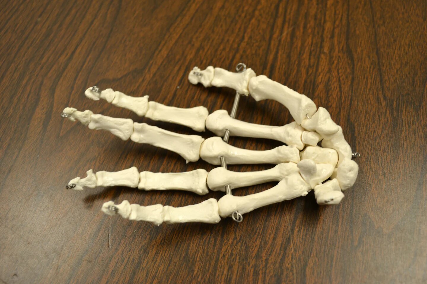
Bi-Lateral Disarticulated Human Skeleton Model features 200+ Bones which includes all of the cuts, marks and grooves needed by every MBBS student.
Anatomy Shop focuses on providing students with the bone sets which have best possible anatomical accuracy so that they can not only score good marks in their medical studies but can also learn deeper concepts of human anatomy.
Each Skeleton Model at AnatomyShop goes through a manual inspection by the team of Experienced Anatomists before being dispatched to customers just to make sure that each skeleton model we dispatch to our customers is anatomically accurate.
Features:
✅[Bi-Lateral Disarticulated Human Skeleton] : As a MBBS student you are required to have deep knowledge under your belt regarding anatomy of human bones and to do so the only option you have is to study the bones in great details for which either you need a real human skeleton which is rare, incomplete, costly and illegal as government of India has banned the trade of "Human Skeletons" so in such a situation the only option you are left with is to get a good quality replica of human bones and our skeleton model does exactly that for you. As it is Bi-Lateral Boneset therefore it comes with both sides of bones and skull with all other tiny bones which are present in human body.
.jpg)
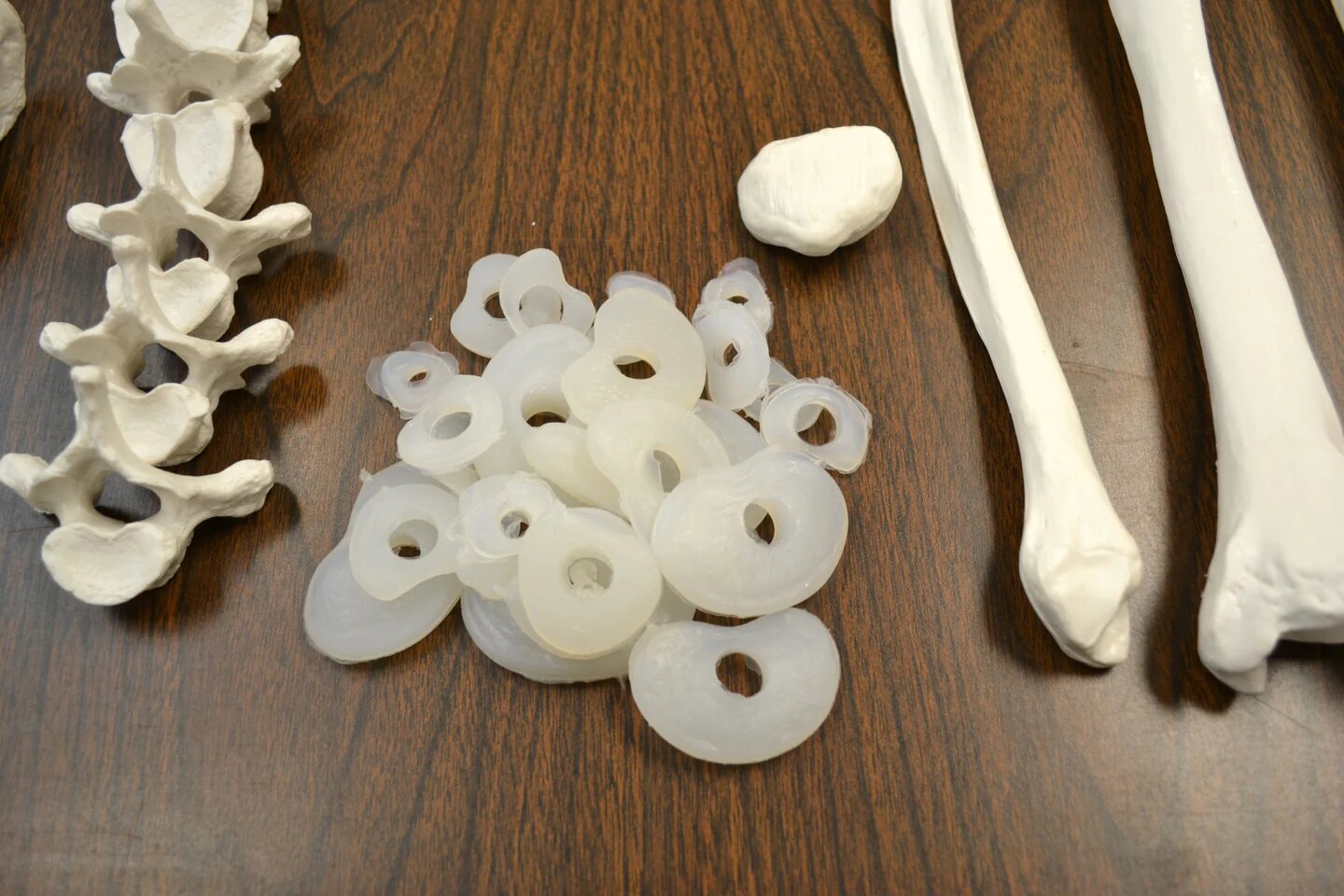
✅[Anatomically Correct] : Bones are casted through the molds which are made from real human specimen which allows our skeleton models to be as precise as real human bones are.
✅ [Robust Quality] : All bones are made from premium quality medical grade pvc plastic which is non-toxic and durable therefore the bones will last for years to come.
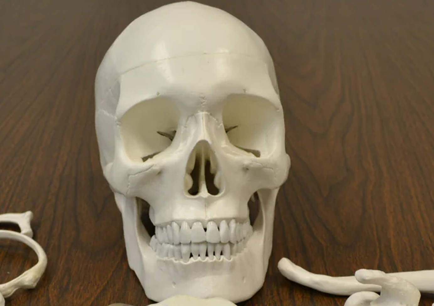
✅[ Premium Skull Model] : The Skull model which comes with this skeleton model can be dissembled into 3 parts for in-depth study of interior and exterior structures, Foramen, fossa, Suture Lines and every tiny anatomical detail is present.
From Textbook to Tabletop: Experience Anatomy in 3D
The 55cm Human Torso Anatomical Model is a professionally crafted, life-representative teaching tool that gives students and educators hands-on access to the human body’s internal systems. Featuring 20 individually removable organs and components—including the brain, lungs, heart, digestive system, and reproductive organs—this model is a must-have for classrooms, clinics, and study environments focused on anatomy, physiology, and health sciences.
At 55cm tall, this unisex anatomical model is compact enough for desks and classrooms but detailed enough for medical-grade instruction. Every organ is designed for accurate representation, intuitive assembly, and educational impact. Whether you're teaching 5th graders about digestion or prepping medical students for practical exams, this model gives you the visual and tactile edge that drives real comprehension.
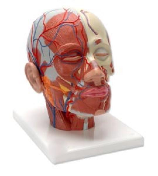
Male Muscle Figure with Internal Organs – 78cm Tall
This life-like Male Muscle Figure with Internal Organs stands 78cm tall and offers a detailed, multi-layered representation of the human muscular and internal organ systems. Ideal for anatomy education and professional demonstration, this model accurately showcases superficial and deep muscles, as well as removable internal organs, allowing for comprehensive study of the human body.
The figure is mounted on a sturdy base and features hand-painted detail for enhanced realism, making it a valuable visual aid in both classroom and clinical settings.
Key Features:
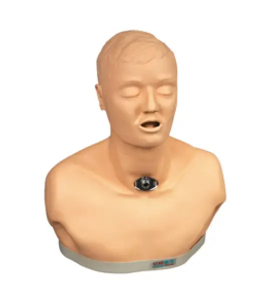
The Adult Tracheotomy Care Manikin is a high-fidelity medical training tool designed to help healthcare professionals and students practice and master the essential skills required for tracheotomy care and airway management. Built with realistic anatomical landmarks and durable materials, this manikin enables effective simulation of routine and emergency tracheostomy procedures, suctioning, and maintenance. It supports hands-on learning and enhances clinical confidence in a controlled, risk-free environment.
Key Features:
This highly detailed Eye with Orbit Model is enlarged 3 times life-size and meticulously crafted to showcase the intricate anatomical structures of the human eye and surrounding orbital region. It is ideal for medical training, patient education, and classroom demonstrations.
The model includes 10 dissectible parts, allowing for hands-on learning and a clear understanding of the eye's anatomy, musculature, and nerve pathways within the orbit. Mounted on a sturdy base, the model offers both durability and visual clarity.
The Giant Eye replica is a great tool to teach-learn the anatomy of the eye! Removable parts of the human eye model include Lens, iris, Vitreous humour, retina
This large anatomical human eye model shows the optic nerve in its natural position in the bony orbit of the eye (floor and medial wall). At three times life size this eye model is great for anatomical demonstrations.
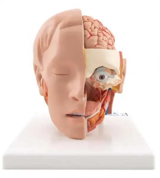
Our most detailed head model! This life-size 6-part head is mounted on a base and features a removable 4-part brain half with arteries.
Representation of the superficial musculature with Parotid gland, Submandibular gland (right half), Deep musculature (left half).
Full size segmented brain features half normal side and three-piece sectioned pathology half, as well as Circle of Willis with aneurism. The brain, which sits inside a partial skull, features the following pathologies which are also illustrated on a two-sided education card: alcoholism, Alzheimer’s, aneurism, depression related tumor, seizure related tumor, migraine, multiple sclerosis, Parkinson’s disease, stroke, and subdural hematoma.
This deluxe brain is medially divided. On the right half of this brain, you will find a colored, systematic grouping and representation of the cerebral lobe.