-
Khanna Traders, are producer and supplier of scientific equipment.
- khannachahat05@gmail.com
-
+91-8930598097
Khanna Traders, are producer and supplier of scientific equipment.
+91-8930598097
With the help of this artery model doctors can explain changes in the blood vessels due to arteriosclerosis.
Anthropological skulls has been newly reissued in its entirety. The results are plain to see in these wonderful anthropolical skulls.
This 3 part medical quality human skull replica is a first choice for basic anatomical studies of the skull. It also makes a great present for medical professionals and students of medicine and allied health professions.
The Physiology of Nerves series displays the basic structures of the human nervous system. Each of the five sections of the nerve model shows a plastic colored relief model of the main synapse variations. All sections of the nerve physiology series can magnetically attach to the illustrated base which depicts the neural components in vivid colors. Each nerve section is also available separately.
This nervous system relief model shows a schematic representation of the central and peripheral nervous system.
The Spinal cord model illustrates the composition of the spinal cord, magnified to a scale of about 5:1. The spinal cord is formed by a central channel surrounded by "gray matter" with an outer layer of "white matter".
This deluxe human torso model is top notch in the field of anatomy. This unique torso depicts both the superficial and deep muscles, and the two main muscles, the deltoid and gluteus maximus can even be removed for closer studies. With this human torso model you can also study the vertebrae, the spinal cord, spinal nerves and vertebral arteries, exchange the male and female genital inserts, discover the internal structures of the brain and much more.
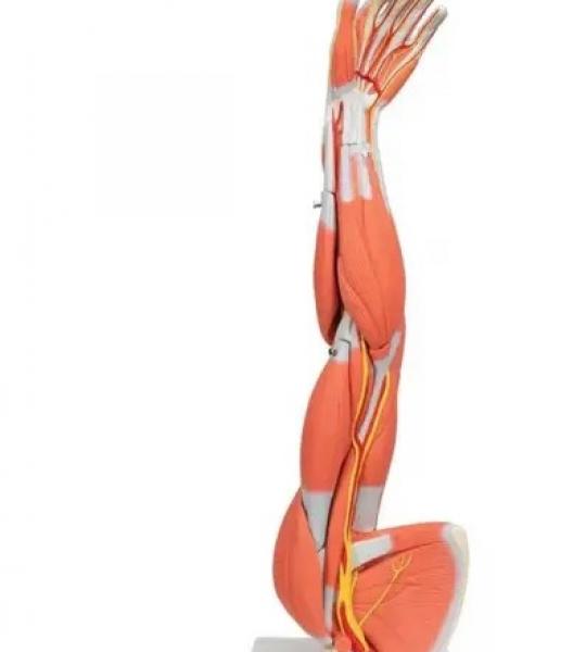
This muscled arm model illustrates both the superficial and deeper muscles, five of which are removable from the muscled arm. Tendons, vessels, nerves and bone components of the left arm and shoulder are shown in great detail on this high quality muscle model.
The foot skeleton features not only the bones but also the muscles, tendons, ligaments, nerves, arteries, and veins of the foot. The frontal view of the foot model features the extensor muscles of the lower leg. The tendons can be followed on their passage under the transverse and crucial crural ligaments all the way to their insertion points.
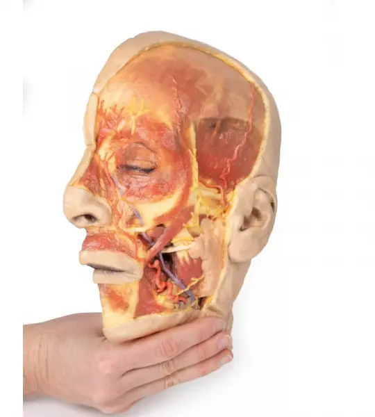
3D model presents a superficial dissection of a left face anterior to the ear with false colouring highlighting a series of neurovascular structures alongside the superficial muscles of facial expression. This compliments the more expanded superficial dissection of the face and lateral head presented in our HW 45 model. The undissected regions of the model have been digitally removed.
Understanding anatomy means seeing how systems interact—and the Human Skeleton Model with Nerves & Blood Vessels gives you that interaction in a compact, accessible format. With clearly marked arteries (red), veins (blue), and peripheral nerves (yellow) mapped across skeletal landmarks, this model offers a detailed multisystem overview that’s easy to move, easy to teach with, and endlessly valuable in both academic and clinical settings.
Standing at approximately 85cm tall (33 inches), this model is ideal for desktop teaching, one-on-one instruction, or visual reinforcement during clinical discussions. It includes major vascular routes and key neural paths that wrap around and pass through a fully articulated skeletal structure. From the carotid arteries and brachial plexus to the femoral nerve and iliac veins, each element is molded with clarity and anatomical precision.
Whether you're a medical student revising for practical exams, a biology teacher explaining human physiology, or a clinician helping patients understand their condition, this model delivers essential insights in a format that fits right on your desk.
Key benefits: